

Background.Cardiovascular magnetic resonance (CMR) is the only available technique for the non-invasive quantification of MIO. The native T1 mapping has recently been proposed as an alternative to the universally adopted T2* technique, due to the higher sensitivity for detection of changes associated with mild or early iron overload.
Objective.To study the association between T1 values and left ventricular (LV) function in thalassemia major (TM) and to evaluate for the first time if T1 measurements quantifying MIO are influenced by macroscopic myocardial fibrosis.
Methods.146 TM patients (87 females, 38.7±11.1 years) consecutively enrolled in the Extension-Myocardial Iron Overload in Thalassemia Network underwent CMR. Native T1 values were obtained by Modified Look-Locker Inversion recovery (MOLLI) sequence in all 16 myocardial segments and the global value was the mean. LV function parameters were quantified by cine images. Late gadolinium enhancement (LGE) technique was used to detect macroscopic myocardial fibrosis.
Results.No correlation was detected between global heart T1 values and LV volume indexes, LV mass index, or LV ejection fraction.
Foourteen (9.6%) patients had an abnormal LV motion (13 hypokinesia and 1 dyskinesia) and they showed significantly lower global heart T1 values than patients without LV motion abnormalities (883.8±139.7 ms vs 959.0±91.3 ms; P=0.049).
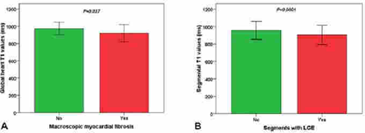
LGE images were acquired in 88 patients (60.3%) and macroscopic myocardial fibrosis was detected in 36 patients (40.9%). The 72.2% of patients had two or more foci of fibrosis. Patients with macroscopic myocardial fibrosis had significantly lower global heart T1 values (921.3±100.3 ms vs 974.5±72.7 ms; P=0.027) (Figure 1A). Data about the LGE was present for 1408 segments (88 patients x 16 segments) and 105 (7.5%) were positive. Segments with LGE had significantly lower T1 values than segments LGE-negative (905.6±110.6 ms vs 956.9±103.8 ms; P<0.0001) (Figure 1B).
Conclusion.No correlation between T1 values and LV function parameters was detected, probably because the majority of the patients had normal or mild abnormal LV parameters. TM patients with macroscopic myocardial fibrosis showed significantly lower T1 values suggesting that T1 measurements for quantifying MIO are not influenced by macroscopic myocardial fibrosis and an association between myocardial iron and macroscopic fibrosis, previously detected only in pediatric TM patients.
Pepe:Chiesi Farmaceutici S.p.A.:Other: no profit support and speakers' honoraria;Bayer:Other: no profit support;ApoPharma Inc.:Other: no profit support.Pistoia:Chiesi Farmaceutici S.p.A.:Other: speakers' honoraria.Meloni:Chiesi Farmaceutici S.p.A.:Other: speakers' honoraria.

This feature is available to Subscribers Only
Sign In or Create an Account Close Modal